Introduction
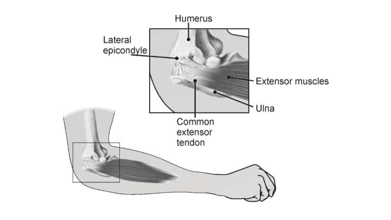
Muscle tissues maintain electrical imbalances, or potentials, across cell membranes by concentrating positive or negative charges on opposite sides of those membranes. These potentials are a form of stored energy. With activation (such as from a nerve impulse), the ions are allowed to cross the muscle cell membranes, generating electrical activity and resulting in muscle contraction.
An electromyogram, or EMG, is a graphical recording of electrical activity within muscles. It is useful in the diagnosis of disorders affecting muscles and the nerves that supply them. Inherited and acquired disorders of muscles (such as the muscular dystrophies), and disorders of the central and peripheral nervous systems (such as Huntington’s disease and diabetic neuropathy) result in abnormal EMG readings.
Objectives
In this experiment, you will
- Obtain graphical representation of the electrical activity of a muscle.
- Associate muscle activity with movement of joints.
- Correlate muscle activity with injury.
Sensors and Equipment
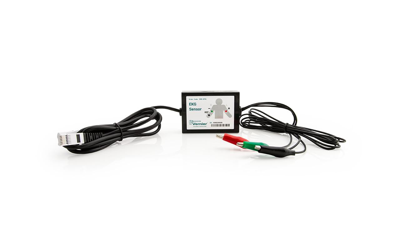
This experiment features the following sensors and equipment. Additional equipment may be required.
Ready to Experiment?
Ask an Expert
Get answers to your questions about how to teach this experiment with our support team.
- Call toll-free: 888-837-6437
- Chat with Us
- Email support@vernier.com
Purchase the Lab Book
This experiment is #15 of Human Physiology with Vernier. The experiment in the book includes student instructions as well as instructor information for set up, helpful hints, and sample graphs and data.


